The systematics of diseases of the oral mucosa is based on the principle of taking into account the etiology and pathogenesis of pathological changes, which makes it possible to develop tactics of treatment and prevention. It should be recognized that this principle is not completely satisfactory due to the fact that in a number of diseases the causes of their occurrence and the mechanism of development are not completely clear.
Based on the requirements of the international classification of diseases as applied to dentistry, Pindborg (I973) recommends the following systematics of diseases of the oral mucosa (in an abbreviated version):
I. Traumatic injuries as a result of mechanical factors
moat, high and low temperatures, electric impact, harmless meteorological
rological factors (meteorological cheilitis, lip cracks), chemical
substances, etc. Form of manifestation: hyperemia, erosion, ulcers, hyperker
toses (leukoplakia).
III.Allergic and toxic-allergic diseases.
1) Contact allergic stomatitis, gingivitis, glossitis, cheilitis (from medicines, plastics and other materials used in dentistry, dyes, toothpastes, elixirs and others chemical substances, ultraviolet rays, etc., in contact with the mucous membrane or red border of the lips).
2) Fixed and widespread toxic-allergic lesions (from medicines, food substances and other allergens that enter the body in various ways).
3) Dermatoses with lesions of the oral mucosa of toxic-allergic genesis (exudative erythema multiforme, Lyell's syndrome, primary systemic vasculitis, including Wegener's syndrome).
IV.Diseases with an autoimmune component of pathogenesis:
1) Recurrent aphthous stomatitis, including cicatricial aphthae;
2) Behcet's syndrome, including Touraine's aphthous disease;
3) Sjogren's syndrome;
4) Dermatoses with lesions of the oral mucosa (pemphigus, pemphigoid, Duhring's disease, lupus erythematosus, scleroderma).
V. Mucocutaneous reaction: lichen planus.
Vi. Changes in the oral mucosa with exogenous intoxication.
Vii. Changes in the oral mucosa and the red border of the lips in the pathology of various organs and systems of the body and metabolic disorders:
1) With visceral and endocrine pathology;
2) With hypo- and avitaminosis;
3) In case of diseases of the blood and blood-forming organs;
4) With pathology of the nervous system;
5) During pregnancy.
VIII. Congenital and genetically determined diseases.
1) Nevi and epithelial dysplasias: vascular nevi, including Sturge-Weber syndrome, verrucous and pigmented nevi, epidermoid cyst, Fordyce's disease, white spongy nevus (mild leukoplakia, "buccal biting", etc.), hereditary benign intraepithelial dyskeratosis.
2) Folded tongue and rhomboid glossitis;
3) Glandular cheilitis;
4) Dermatoses with lesions of the mucous membrane of the oral cavity and lips: bullous
epidermolysis, atopic dermatitis (cheilitis), psoriasis, ichthyosis,
darier's disease, Peutz-Jeghers-Touraine syndrome, congenital pachyonychia,
anhydrotic epithelial dysplasia.
IX. Precancerous diseases, benign and malignant neoplasms.
1) Obligate precancer: Bowen's disease, verrucous precancer, limited
ny hyperkeratosis of the red border of the lips, abrasive precancerous hey
lit. Manganotti.
2) Optional precancer: leukoplakia, keratinizing papilloma and pa-
pillomatosis, keratoacanthoma, cutaneous horn, etc.
3) Benign neoplasms.
4) Cancer.
The given systematization in the future, with the accumulation of knowledge about the etiology and pathogenesis of diseases of the oral mucosa and lips, should be improved.
The classification of diseases and lesions of the oral mucosa in childhood was proposed by T.F. Vinogradova et al. (1974).
I. According to the clinical course: acute and chronic (recurrent and per
manent).
II. Clinically pronounced morphological changes: primary
changes (catarrhal, fibrinous, alterative and proliferative
inflammation; vesicular, blistering and papular rashes), secondary
changes (erosion, aphthae, ulcer).
III. By localization: papillitis, gingivitis, pareitis, glossitis, palatinitis, stomatitis. IV. By etiology:
1.Injuries resulting from mechanical, physical and chemical injury
(abrasion, decubital ulcer, Bednar's aphthae, leukoplakia, chronic tre
lip, radiation, chemical and thermal burns, actinic hey
litas, gingivitis due to abnormal attachment of the frenum, etc.).
2.Diseases caused by:
- viral infections (herpetic stomatitis, enterovirus stomatitis, herpangina, foot and mouth disease, vesicular stomatitis, measles stomatitis, chickenpox, etc.);
- bacterial infections (gonorrheal stomatitis, tuberculous stomatitis, glossitis, cheilitis), staphylococcal pyoderma, etc.;
- fungal infections (acute superficial candidiasis - thrush, yeast glossitis, candidomycotic seizure, deep candidiasis, actinomycosis, etc.), spirochete and fusospirochete (syphilis, Vincent's stomatitis, etc.);
3. Diseases caused by allergic reactions in contact, microbial and drug allergies (erythema multiforme, Stevens-Johnson, Fissenzhe-Randu, Lyell, Reiter syndromes, chronic recurrent aphthous stomatitis, etc.).
4.Changes and diseases of the oral mucosa, which are symptoms or manifestations of the pathology of other organs and systems of the body and arising from:
- blood diseases (desquamative glossitis - "Hunter's language" with anemia, ulcerative stomatitis with reticulosis: acute and chronic leukemia, lymphogranulomatosis, neutropenia, etc.);
- skin diseases (lichen planus, Duhring's dermatitis, epidermolysis bullosa, etc.);
- diseases of the gastrointestinal tract and liver (acute catarrhal and ulcerative stomatitis with dysentery, chronic recurrent aphthous and ulcerative stomatitis, Setton's stomatitis, desquamative glossitis, diamond-shaped glossitis, chronic catarrhal, hypertrophic and ulcerative gingivitis and stomatitis, etc.);
-sharp infectious diseases (Filatov-Koplik spots with measles, raspberry tongue with scarlet fever, vesicular stomatitis with chickenpox, ulcerative necrotizing stomatitis with typhoid fever, hemorrhages and increased vascular pattern with influenza, catarrhal stomatitis with pronounced granular mucous membrane with adenovirus infection, etc.);
- systemic diseases (lupus erythematosus, Wegener's syndrome, eosinophilic collagenosis, reticulohistiocytosis - Hand-Schüller-Christian disease, etc.);
- cardiovascular diseases (Osler's disease, chronic catarrhal gingivitis and stomatitis with blue heart disease, chronic ulcerative stomatitis, etc.);
-endocrinopathies (hypertrophy and folding of the tongue in Down's disease, scrotal tongue in Shereshevsky-Turner syndrome, generalized periodontal disease in diabetes, periodontopathy in dyshormonal endocrinopathies, etc.);
- neuropsychiatric diseases (gingivitis with oligophrenia, mild leukoplakia, rhomboid and desquamative glossitis, etc.).
Particular attention should be paid to the advisability of isolating such etiological factors that directly cause diseases, damage or lesions of the oral mucosa.
The elimination of these causes should entail the elimination of the pathology. Along with this, diseases of systems and organs act as the cause, in which changes and lesions of the oral mucosa are a symptom of a disease or a reflection of the general patterns of a pathological process in the body.
In these situations, the doctor's tactics in the diagnosis and treatment of a child should be comprehensive, using the methods and knowledge of doctors of the relevant profiles. The effectiveness of treatment depends on the child's health status and the success of therapy common disease... The role of local treatment of stomatitis, despite its advisability, is only of secondary importance.
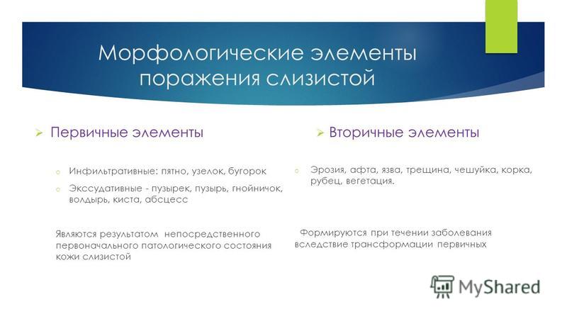
Elements of the lesion can be primary and secondary (developed from primary).
Diseases of the oral mucosa (OOM), arising in isolation as a result of the action of various factors on the oral mucosa, account for 10 to 15% of all pathological lesions of the oral mucosa. Most of the diseases of the oral mucosa are syndromes of various genesis of diseases of organs or body systems (gastrointestinal tract, endocrine system, and others).
Currently, there are several classifications of diseases of the oral mucosa. Classifications of A.I. Rybakov and E.V. Borovsky et al. Were and are widespread in Russia. The official is the WHO nomenclature (ICD-X, Geneva):
K 12. Stomatitis and related lesions / excluded:Herpetic sore throat (08.5K); Stomatitis: acute gangrenous (A 69.0), allergic (L 23.0), candidal (B 37.0), contact (K.12.14), caused by the Coxsackie virus, NOS (B.34.1), drug (T 36-T 50), mycotic ( B.37.0), nicotine (K.13.24.), Vesicular (B.08.4.) And others /;
K 12.0. Recurrent aphthous oral cavity (Aphthous stomatitis; Ulcerative lesion; Mikulich's aphthae; Small aphthae; Recurrent aphthous ulcers);
K 12.01. Recurrent muco-necrotic periadenitis: Cicatricial aphthous stomatitis; Large aphthae; Afta Satten;
By 12.02. Herpetiform stomatitis;
By 12.03. Afty Bednar;
By 12.04. Traumatic ulceration (including prosthetic);
By 12.08. Other specified recurrent oral aphthae;
By 12.09. Recurrent oral aphthae, unspecified;
K 12.1. Other forms of stomatitis: By 12.10. Artificial stomatitis; By 12.11. "Geographic" stomatitis; By 12.12. Prosthetic stomatitis / excluded: Candidal stomatitis associated with the use of a prosthesis (В 37.03.) /;
By 12.13. Papillary hyperplasia of the palate;
By 12.14. Contact stomatitis - "cotton roll" stomatitis;
By 12.18. Other specified forms of stomatitis;
By 12.19. Stomatitis, unspecified;
K 13. Other diseases of the lips and oral mucosa:
K 13.0. Diseases of the lips:
By 13.00. Angular cheilitis, angular cheilosis, seizure (crack, lip adhesion);
By 13.01. Glandular cheilitis;
By 13.02. Cheilitis exfoliative;
By 13.03. Heilit NOS;
By 13.04. Helodynia;
By 13.08. Other specified lip diseases
By 13.09. Diseases of the lips, unspecified;
K 13.1. Biting the cheek and lips;
By 13..2. Leukoplakia and other changes in the epithelium of the oral cavity, including the tongue;
By 13.20. Leukoplakia is idiopathic;
By 13.21. Lekoplakia nicotine;
By 13.22. Erythroplakia;
By 13.23. Aikedema;
By 13.24. Smoker's palate (nicotine leukokeratosis; nicotine stomatitis);
By 13.28. Dr. changes in the epithelium;
By 13.29. Unspecified epithelial changes, leukoplakia NOS;
K 13.3. Hairy lekoplakia;
To 13.4. Granuloma and granulomatous lesions of the oral mucosa:
By 13.40. Pyogenic granuloma;
By 13.41. Eosinophilic granuloma of the oral mucosa;
By 13.42. Verrucous xanthoma (histiocytosis Y);
By 13.48. Other specified granulomas;
By 13.49. Unspecified granulomas;
To 13.5. Submucous fibrosis of the oral cavity;
To 13.6. Hyperplasia of the oral mucosa due to irritation / excluded: Hyperplasia of the edentulous alveolar edge due to irritation (hyperplasia associated with wearing a prosthesis - K.06.23);
K 13.7. Other and unspecified lesions of the oral mucosa:
By 13.70. Excessive melanin pigmentation (melanoplakia, smoker's melanosis);
By 13.71. Oral fistulas;
By 13.72. Voluntary tattoo;
By 13.73. Focal mucinosis of the oral cavity;
By 13.78. Dr. specified lesions of the oral mucosa;
By 13.79. Unspecified lesion of the oral mucosa;
K 14. Diseases of the tongue (excluded: congenital macroglossia - G 38.2x);
K 14.0. Glossitis
By 14.01. Traumatic ulceration of the tongue;
By 14.08. Dr. specified glossitis;
By 14.09. Glossitis, unspecified;
K 14.1. "Geographic" language (Benign migratory glossitis, Exfoliative glossitis);
K 14.2. Median rhomboid glossitis;
K 14.3. Hypertrophy of the papillae of the tongue;
By 14.30. Coated tongue;
By 14.31. "Hairy" tongue, Black "hairy" tongue; Black villous tongue;
By 14.32. Dr. specified hypertrophy of the papillae of the tongue (hairy tongue due to taking antibiotics);
By 14.39. Hypertrophy of the papillae of the tongue, unspecified;
K 14.4. Atrophy of the papillae of the tongue
By 14.40. caused by the habits of clearing the tongue;
By 14.41. caused by a systemic disorder;
By 14.42. Atrophic glossitis NOS;
By 14.48. Dr. specified atrophy of the papillae of the tongue;
By 14.49. Atrophy of papillae of tongue, unspecified;
K 14.5. Folded tongue (wrinkled, furrowed, split);
K 14.6. Glossodyne;
By 14.60. Glossopyrosis (burning sensation in the tongue);
By 14.61. Glossadinia (pain in the tongue);
By 14.68. Other specified glossadinia;
By 14.69. Glossadinia, unspecified;
K 14.8. Other diseases of the tongue:
By 14.80. Serrated tongue (with teeth marks);
By 14.81. Hypertrophy of the tongue;
By 14.82. Tongue atrophy;
By 14.88. Other specified diseases of the tongue;
By 14.9. Diseases of the tongue, unspecified.
We believe that the classification of A.I. Rybakov, like the WHO, based on the nosological, and not on the etiological principle of construction, is more convenient for teaching this section. Therefore, the presentation of the material will correspond to the structure of these classifications.
The classification of diseases and lesions of the oral mucosa in childhood has its own characteristics. Currently, two classifications are widespread - T.F. Vinogradova with co-authors (1974) and V.V. Zhilina (1991). T.F. Vinogradova et al (1974) proposed to systematize diseases of the oral mucosa as follows:
- by clinical course:acute and chronic (recurrent and permanent);
- by morphological changes:primary changes (catarrhal, fibrinous, alterative and proliferative inflammation; vesicular, cystic and papular rashes), secondary changes (erosion, aphthae, ulcer);
- by localization:papillitis, gingivitis, pareitis, glossitis, palatinitis, stomatitis;
- by etiology:
a) damage as a result of mechanical, physical and chemical trauma (abrasion, decubital ulcer, Bednar's aphthae, leukoplakia, chronic lip fissure, radiation, chemical and thermal burns, actinic cheilitis, gingivitis caused by abnormal attachment of the frenum, etc.);
b) diseases caused by:
Viral infections (herpetic stomatitis, enterovirus stomatitis, herpangina, foot and mouth disease, vesicular stomatitis, measles stomatitis, chickenpox, etc.);
Bacterial infections (gonorrheal stomatitis, tuberculous stomatitis, glossitis, cheilitis), staphylococcal pyoderma, etc.;
Fungal infections (acute superficial candidiasis - thrush, yeast glossitis, candidomycotic seizure, deep candidiasis, actinomycosis, etc.), spirochete and fusospirochete (syphilis, Vincent's stomatitis, etc.);
c) diseases caused by allergic reactions in contact, microbial and drug allergies (exudative erythema multiforme, Stevens-Johnson, Fissenzhe-Randu, Layel, Reiter's syndromes, chronic recurrent aphthous stomatitis, etc.);
d) changes and diseases of the oral mucosa, which are symptoms or manifestations of the pathology of other organs and systems of the body and arising from:
Diseases of the blood (desquamative glossitis with anemia, ulcerative stomatitis with reticulosis, acute and chronic leukemia, lymphogranulomatosis, neutropenia, etc.);
Diseases of the skin (lichen planus, Duhring's dermatitis, epidermolysis bullosa, etc.);
Diseases of the gastrointestinal tract and liver (acute catarrhal and ulcerative stomatitis in dysentery, chronic recurrent aphthous and ulcerative stomatitis, Setton's stomatitis, desquamative glossitis, diamond-shaped glossitis, chronic catarrhal, hypertrophic and ulcerative gingivitis and stomatitis, etc.);
Acute infectious diseases (Filatov-Koplik spots with measles, raspberry tongue with scarlet fever, vesicular stomatitis with chickenpox, ulcerative-necrotizing stomatitis with typhoid fever, hemorrhages and increased vascular pattern with influenza, catarrhal stomatitis with pronounced granular mucous membrane with adenovirus infection and other .);
Systemic diseases (lupus erythematosus, Wegener's syndrome, eosinophilic collagenosis, reticulohistiocytosis - Hend-Schüller-Christian disease, etc.);
Cardiovascular diseases (Osler's disease, chronic catarrhal gingivitis and stomatitis with blue heart disease, chronic ulcerative stomatitis, etc.);
Endocrinopathies (hypertrophy and folding of the tongue in Down's disease, scrotal tongue in Shereshevsky-Turner syndrome, generalized periodontal disease in diabetes, periodontal disease in dyshormonal endocrinopathies, etc.);
Neuropsychiatric diseases (gingivitis in oligophrenia, mild leukoplakia, rhomboid and desquamative glossitis, etc.).
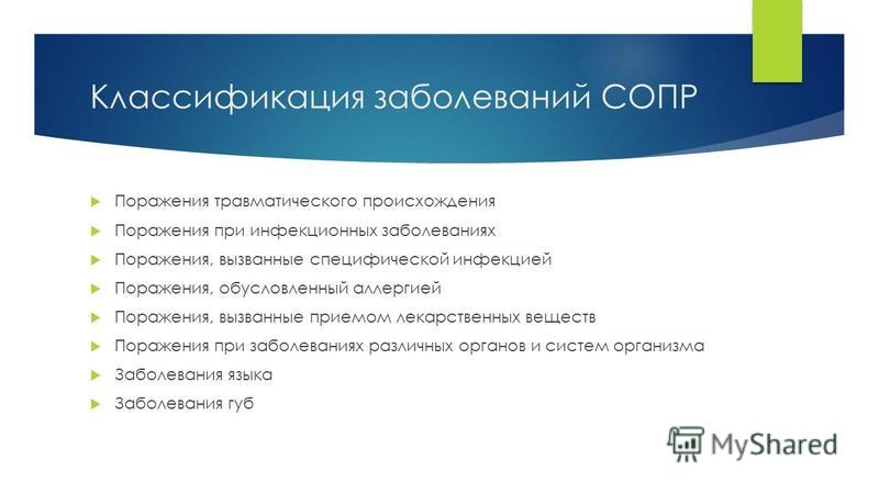
V.V. Zhilina (1991) proposed another classification of diseases of the oral mucosa, tongue and lips in children (The authors consider this classification to be the most successful for a practical dentist, therefore, the peculiarities of the course of diseases of the oral mucosa in children were presented in accordance with it. Approx. editors):
1) Lesions of the oral mucosa of traumatic origin.
2) Lesions of the oral mucosa in infectious diseases.
3) Lesions of the oral mucosa caused by a specific infection.
4) Lesions of the oral mucosa caused by allergies.
5) Lesions of the oral mucosa caused by the intake of medicinal substances.
6) Changes in the oral mucosa with diseases of various organs and systems of the body.
7) Diseases of the tongue.
8) Diseases of the lips.
Pathological processes of various origins alter the metabolism of the oral mucosa and in most cases are accompanied by its structural changes. The processes occurring in the epithelium and the lamina propria are classified as reactive,or destructive.
The former include acanthosis, papillomatosis, parakeratosis, and grade I dysplasia. Reactive changes are most often inflammatory. In cases acute inflammation, flowing in a short period of time, exudation processes prevail. In other cases, they may be the result of degenerative changes in the mucous membrane.
Due to the anatomical features of the structure of the oral mucosa, edema and hyperemia are not always clearly defined in the pathology of the oral mucosa. Most often, inflammation is manifested by desquamation of the epithelium or the appearance of cavity formations with serous or hemorrhagic exudate - a vesicle, a bladder. In a chronic reaction, symptoms of inflammation such as exudation are less pronounced, the infiltrative component of inflammation prevails with the proliferation of cellular elements. Therefore, primary ejection elements are subdivided into exudative and infiltrative (see below).In addition, primary exudative elements are rapidly transformed into secondary (see below).
Primary pouring elements:
exudative:
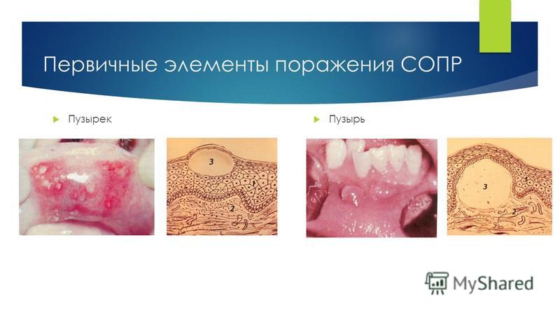
- bubble(vesicula) - endothelial cavity formation on an unchanged or hyperemic mucous membrane, with more serous contents;
- bubble(bulla) - a cavity element of a larger size, located intra- or subepithelially, with serous or hemorrhagic contents;
infiltrative:
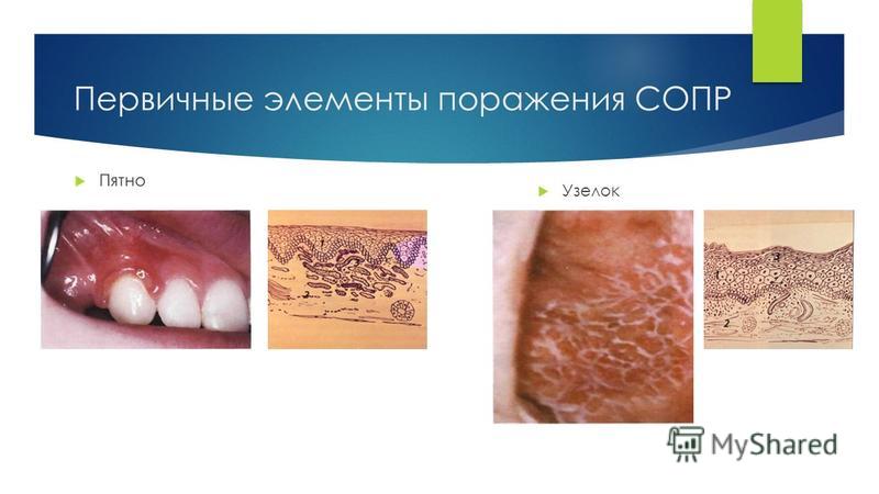
- spot(macula) - a limited area of \u200b\u200bthe mucous membrane changed in color due to venous or arterial hyperemia. Inflammatory spots are observed, for example, with erythema multiforme exudative. Non-inflammatory spots arise as a result of the deposition of dyes of endo- and exogenous origin (pigmentation in jaundice, when exposed to occupational hazards, etc.);
- nodule(papula) is a noncavity infiltrative formation ranging in size from a pinhead to 10 mm in diameter, slightly rising above the level of the mucous membrane. The process is localized in the epithelium: acanthosis, a granular layer appears, hyperkeratosis, parakeratosis. After the nodule has healed, there are usually no scars. An example is lichen planus;
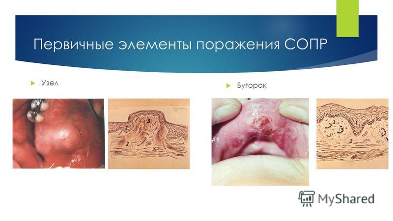
- tubercle(tuberculum) is a noncavity infiltrative formation that rises above the level of the mucous membrane. The infiltrate captures all layers of the mucous membrane, quickly disintegrates and leaves a scar after the resolution of the process. An example of it is tuberculous lupus, tubercular syphilis;
- knot(nodus) is a large oval formation located in all layers of the mucous membrane. The node either rises above the level of the mucous membrane, or is felt in its thickness. With suppuration, fistulas may form. Leaves a scar after resolution. An example is leprosy, tertiary syphilis.
Secondary discharge elements:
flake(squama) - with incomplete keratinization - parakeratosis of the mucous membrane, scales appear. They are defined as mica translucent plates fixed in the middle to the red border of the lips. An example is exfoliative cheilitis;
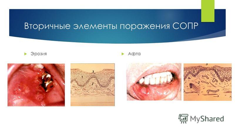
erosion(erosio) - defect of the mucous membrane within the epithelium. Heals erosion without scarring. An example is erosion in herpetic stomatitis;
excoriation(excoriatio) - abrasion, traumatic erosion, damage to the deeper layers of the epithelium up to the papillary layer;
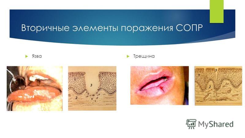
ulcer(ulcus) - a defect of the mucous membrane, capturing its own layer of the mucous membrane or deeper tissues. The bottom and edges of the ulcer are of a different nature. After healing, the ulcer leaves a scar, as for example, with a disintegrated gum, tuberculous ulcer, Setton's aphtha;
crack(rhogas) - a linear defect of the mucous membrane. An example is a jam, a crack in the red border of the lips;
aphtha(aphta) - an oval-shaped epithelial defect with clear boundaries. At the bottom there is a dense fibrinous plaque. An example is chronic aphthous stomatitis;
crust(crusta) - is formed due to drying on the mucous membrane, more often the red border of serous exudate or blood;
scar(cicatrix) - represents the replacement of destroyed tissues with connective tissue. It is structurally and functionally defective tissue. Scars are dense and soft in consistency. Soft thinned, slightly sinking scars are called atrophic;
vegetation -overgrowth of papillae of the mucous membrane's own milk on the surface, erosion. An example is papillary hyperplasia of the mucous membrane as a result of chronic injury with a prosthesis;
pigmentation -arises on the basis of previous inflammatory changes, in which there was a hemorrhage in the tissue with a subsequent change in color in accordance with the shades that the dyes of the blood take;
lichenization(lichenisatio) - thickening of the mucous membrane with the fusion of papular elements, for example, with lichen planus.
With a thorough examination of patients with diseases of the mucous membrane, it is extremely important to find out concomitant somatic diseases, occupational hazards, and possible mental disorders.
Inflammation Is a protective and adaptive response to a spherical-horny stimulus. The development of inflammation, the forms of its manifestation, the nature of the course are smallpox signs that are usually taken into account when constructing systematics or classifications of diseases of the oral mucosa.
By the nature of the flow inflammatory process can develop as an acute, subacute, chronic or exacerbated. Natakis groups usually divide diseases in taxonomists.
The acute course of inflammation, the localization of the process on the oral mucosa, the peculiarities of the clinical signs of the disease most fully reveal its sugary. This characteristic is usually reflected in its name (for example, acute herpetic stomatitis; acute ulcerative gingivitis, etc.). The main manifestation of the acute course of the disease is the vascular-exudative reaction with the prevalence of alteration processes. In chronic inflammation against the background of edema and infiltration, proliferative phenomena with varying degrees of proliferation of fibrous connective tissue structures are predominant. With an exacerbation of the flow chronic inflammation against the background of changes natural for chronic inflammation, vascular permeability increases, exudation and alteration phenomena increase, and polymorphonuclear leukocytes appear in the infiltrate.
This course is observed in certain forms of cheilitis, glossitis, gangivitis and other diseases. This development is especially often observed in persons with diseases of the gastrointestinal tract, endocrine organs, blood, and with radioactive irradiation.
Another, no less important factor, which is often taken into account when classifying, is the form of inflammation: exudative, alterative or protractive. With each disease, some form of inflammation prevails. At the same time, the development of various forms of inflammation with dystrophic disorders (keratosis) or the combination of inflammation and dystrophy, the formation of scar tissue (lupus erythematosus) is observed.
In the acute course, most stomatitis develops with the advantage of exudative processes - catarrhal stomatitis, cheilitis, medicinal stomatitis, radiation mucositis, etc. With the development of these processes, following a short-term reflex narrowing of the lumen of all sections of superficially located small vessels, their persistent expansion occurs, which leads to a slowdown in blood flow and stasis. Persistent hypoxia develops in the affected area of \u200b\u200bthe oral mucosa.
Deeper changes in many diseases of OCPD occur with the altsratin form of inflammation. Clinically, with this course, there are limited or extensive violations of the integrity of the integumentary epithelium, and often the own plate of CO. Vivid examples of such development are recurrent aphthous stomatitis, exudative erythema multiforme, Vincent's stomatitis, traumatic or specific ulcer, etc. In these diseases, inflammation and dystrophy are observed, which accompany the gay with necrotic processes, covering cellular elements, fibrous structures and the main substance of the oral mucosa. Deep changes occur in the vessels, there are signs of a reaction of the surrounding tissues, lymphadenitis, etc.
Sagging of the oral mucosa, in which proliferative inflammation prevails, is characteristic of hypertrophic papillitis, gingivitis, warty (verrucous) form of leukoplakia. The basis of morphological changes in these diseases is the multiplication and transformation of cellular structures leading to the formation and proliferation of connective tissue structures, the formation of granulation tissue, the development of granulomas.
In the early stages of the study of the pathology of oral mucosa in the systematization of diseases of the mucous membrane, mainly approaches based on clinical signs of the disease were used. In such a class of diseases, diseases were combined into separate groups according to a common symptom characteristic of them. For example, if among the clinical signs of the disease the main element was a bladder or vesicle, they were combined into a group 1 of cystic stomatitis (herpetic stomatitis, Duhring's disease, pemphigus, etc.). If the leading was pain syndrome (neuralgia of the trigeminal, glossopharyngeal nerves, damage to the ganglia, glossodynia), such diseases were combined into a group of neurogenic lesions. With the simultaneous loss of the process on the OSR and the skin, the diseases were combined into a group of dermatostomatitis. This approach to assessing the manifestations of the disease was of great importance for the choice of symptomatic treatment methods.
Summarizing the materials that illuminate different aspects of the pathological processes occurring in the OSS, it should be emphasized that those published over the past 40-50 years in domestic and foreign literature, numerous from the classification have made a certain contribution to the system of studying and understanding this section, which is important for practical dentistry. Of particular interest are those classifications in the structure of which, to one degree or another, the main etiological, pathogenetic and clinical signs of individual diseases are reflected. And it is not the researchers' fault that there is still no comprehensive classification that would clearly reveal the essence of each of the diseases, which would make it possible to construct a method of its treatment in the most professional way and outline rational nougats of prevention.
Difficulties in creating an optimal variant of the systematics of diseases of the oral mucosa and adjacent skin I juices are due to a variety of etiological, pathogenetic and clinical signs, which naturally combine with the development of a certain disease. This, first of all, explains the existence of classifications of different methodological approaches, which are used by practitioners of I in different regions of the former USSR.
In conclusion, we consider it advisable to cite the most common classifications of diseases of oral mucosa, which were set out in textbooks published in the former USSR.
One of the earliest taxonomists of CO diseases is the classification by I.G. Lukomskoy (1945).
1. The main group of stomatitis:
a) superficial - catarrhal, aphthous candidiasis, leukoplakia;
b) deep - ulcerative, hanfenous, hypertrophic.
2. Symptomatic stomatitis - with infectious diseases, blood and metabolic diseases.
3. Dermatostomatitis:
a) stomatitis with symptoms of hyperkeratosis;
b) stomatitis with blistering and vesicular phenomena.
4. Specific stomatitis - syphilis, tuberculosis, leprosy.
5. Isolated lesions of the tongue and lips.
A.I. Rybakov's classification (1964), into which in 1978 clinically and experimentally substantiated additions by A.I. Rybakov and G.V. Banchenko were made:
Stomatitis: catarrhal, acute aphthous (mild, moderate, severe), including solitary aphthae, labial herpes, necrotic ulcers, chronic recurrent aphthous (fibrinous, necrotic, scarring, deforming, glandular, lichenoid forms) ulcerative-necrotic.
Gingivitis:catarrhal, ulcerative-necrotic, hypertrophic, atrophic, desquamative, symptomatic.
Diseases of the tongue: acute inflammatory (catarrhal glossitis, ulcerative, desquamative, tongue abscess), chronic (geographical tongue, villous, diamond-shaped, folded), tongue systemic diseases, tongue anomalies.
Diseases of the lips (cheilitis: catarrhal, glandular, exfoliative, eczematous, meteorological; chronic lip cracks).
Damage and allergic lesions of the OAS: mechanical, physical, chemical, medicinal, professional, prosthetic stomatitis, allergic reactions.
Lesions of the oral mucosa in diseases of internal organs, infectious diseases, specific infections, hypovitaminosis.
ODS candidiasis.
Lesions of oral mucosa in dermatoses (multiforme exudative erythema, pemphigus, lichen planus, lupus erythematosus, etc.).
Pretumor and neoplastic lesions: Talpeiner's leukoplakia, flat, verrucous, nevi oral cavity , abrasive precancerous cheilitis of Manganotti, limited precancerous hyperkeratosis of the lower lip, warty precancer of the red border of the lower lip, Bowen's disease and Keir's erythroplasia, cutaneous horn, cancer and other tumors.
Manifestations of syndromes on oral mucosa.
A slightly different classification is used at the Moscow Medical Dental Institute (MMOMA). The MMSI classification (1972) includes 9 groups of diseases. It should be noted that separate changes and additions were made to different editions that published this classification. The most significant of them are contained in the "Atlas of OCD diseases" (E.V. Borovsky, N.F. Danilevsky, 1981, 1982), where this classification is given by two authors - E.V. Borovsky and A.L. Mashkillyson.
Classification of diseases of the oral mucosa. Etiology. Pathogenesis. Traumatic lesions of the OAS. DEPARTMENT OF CHILDREN'S DENTAL AND ORTHODONTICS Completed by students of the Faculty of Dentistry of the 5th course State educational institution of higher professional education First Moscow State Medical University named after I.M. Sechenov
Classification of diseases of oral mucosa Lesions of traumatic origin Lesions in infectious diseases Lesions caused by a specific infection Lesions caused by allergy Lesions caused by the intake of medicinal substances Lesions in diseases of various organs and systems of the body Diseases of the tongue Diseases of the lips
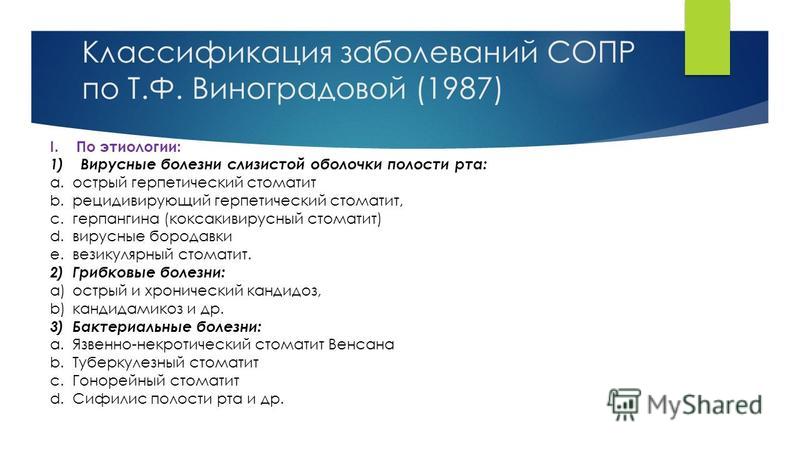
Classification of diseases of oral mucosa according to T.F. Vinogradova (1987) I. By etiology: 1) Viral diseases of the oral mucosa: a. Acute herpetic stomatitis b. Recurrent herpetic stomatitis, c. Herpangina (coxacivirus stomatitis) d. Viral warts e. Vesicular stomatitis. 2) Fungal diseases: a) acute and chronic candidiasis, b) candidiasis, etc. 3) Bacterial diseases: a. Necrotizing ulcerative stomatitis of Vincent b. Tuberculous stomatitis c. Gonorrheal stomatitis d. Syphilis of the oral cavity, etc.
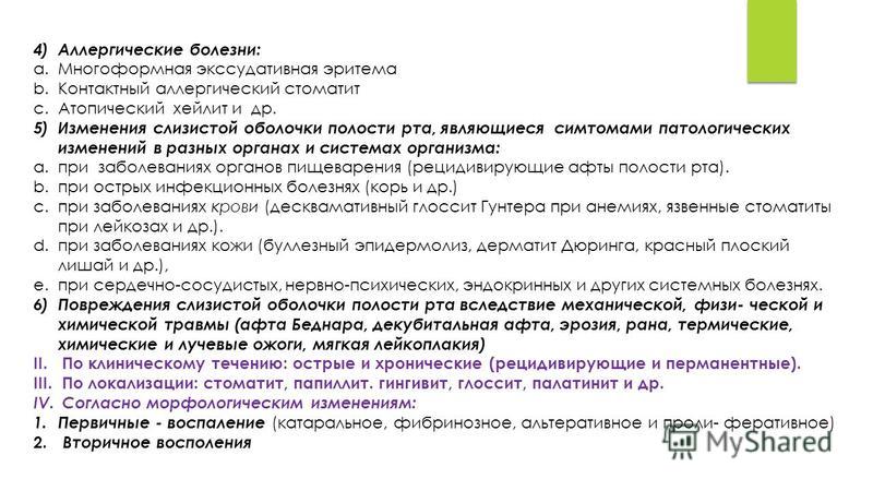
4) Allergic diseases: a. Exudative erythema multiforme b. Contact allergic stomatitis c. Atopic cheilitis, etc. 5) Changes in the oral mucosa, which are symptoms of pathological changes in various organs and systems of the body: a. In diseases of the digestive system (recurrent aphthae oral cavity). b. in acute infectious diseases (measles, etc.) c. for blood diseases (Gunther's desquamative glossitis with anemia, ulcerative stomatitis with leukemia, etc.). d. for skin diseases (epidermolysis bullosa, Duhring's dermatitis, lichen planus, etc.), e. for cardiovascular, neuropsychic, endocrine and other systemic diseases. 6) Damage to the oral mucosa due to mechanical, physical and chemical trauma (Bednar's aphthae, decubital aphthae, erosion, wound, thermal, chemical and radiation burns, mild leukoplakia) II. According to the clinical course: acute and chronic (recurrent and permanent). III. By localization: stomatitis, papillitis. gingivitis, glossitis, palatinitis, etc. IV. According to morphological changes: 1. Primary - inflammation (catarrhal, fibrinous, alterative and proliferative) 2. Secondary inflammation
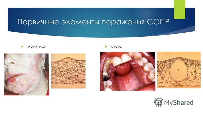
Morphological elements of mucosal lesions Primary elements o Infiltrative: spot, nodule, tubercle o Exudative - vesicle, bladder, abscess, blister, cyst, abscess Are the result of the immediate initial pathological condition of the mucosal skin Secondary elements o Erosion, aphthae, ulcer, crack, scale, crust , scar, vegetation. Formed during the course of the disease due to the transformation of primary
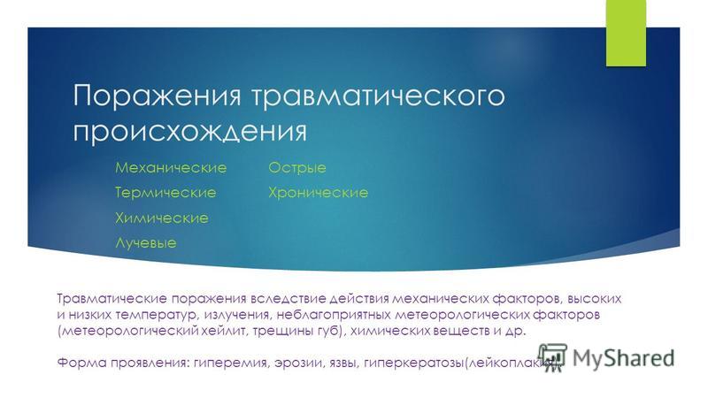
Lesions of traumatic origin Mechanical Thermal Chemical Radiation Acute Chronic Traumatic injuries due to mechanical factors, high and low temperatures, radiation, adverse meteorological factors (meteorological cheilitis, cracked lips), chemicals, etc. Form of manifestation: hyperemia, erosion, ulcers, hyperkeratosis ( leukoplakia).
Therapeutic dentistry. Textbook Evgeny Vlasovich Borovsky
11.1. CLASSIFICATION OF DISEASES OF THE ORAL MUCOSA
Difficulties in diagnosing and choosing a method of treatment for pathology of the oral mucosa are due to the variety of manifestations of diseases localized in this area and often having similar clinical manifestations with different etiology or pathogenesis. The classification of diseases of the oral mucosa is very important for the practitioner, since it helps to navigate their existing variety, thereby contributing to the correct diagnosis, the choice of justified treatment methods and preventive measures.
Previous classifications of diseases of the oral mucosa were based on different principles: anatomical and clinical (stomatitis, cheilitis, glossitis), the nature of the course (acute and chronic), clinical and morphological signs (catarrhal, ulcerative, etc.), the depth of the lesion (superficial and deep ), the nature of the rash (bullous, ulcerative, vesicular, etc.). However, each of them had its own shortcomings and did not fully meet the above classification requirements.
The World Health Organization (WHO) has adopted the following nomenclature of diseases:
II. Neoplasms
III. Diseases of the blood and blood-forming organs and lesions caused by the immune mechanism
IV. Endocrine, metabolic and nutritional diseases
V. Mental and character disorders
Vi. Diseases of the nervous system
IX. Diseases of the circulatory system
X. Diseases of the respiratory system
XI. Diseases of the digestive system
XII. Diseases of the skin and subcutaneous tissue
XIII. Diseases of the musculoskeletal system and connective tissue
XIV. Diseases of the genitourinary system
XV. Pregnancy, childbirth and the postpartum period
XVII. Congenital defects, deformities and chromosomal abnormalities
Xviii. Symptoms, signs, abnormalities, not elsewhere classified
XIX Injuries, poisoning and other consequences of external factors
XX. External factors leading to morbidity and mortality
For each of the listed sections of the classifications, there is a list of dental diseases and syndromes manifested in the oral cavity, characteristic of a given systemic pathology or condition. Systematization of dental diseases within a group is carried out according to clinical, morphological, pathomorphological signs, the nature of microflora, etc.
The main goal of creating an international nomenclature of dental diseases and syndromes in the oral cavity is to standardize approaches to identifying dental pathology with subsequent registration and determination of the level of dental morbidity in different countries. In addition, the information obtained allowed us to determine the correlations of the general state of the body with the level of dental health, as well as to identify systemic diseases associated with pathology in the oral cavity.
Thus, at present, the etiological and pathogenetic relationship between diseases of various organs and systems and pathologies in the oral cavity has been proven, and primarily diseases of the oral mucosa.
To systematize the known pathological conditions (diseases) of the oral mucosa, E.V. Borovsky and A.L. Mashkillyson (1984) proposed to group them, taking as a basis etiological or pathogenetic factor, in the following way.
I. Traumatic lesions due to the action of mechanical factors, high and low temperatures, radiation, unfavorable meteorological factors (meteorological cheilitis, cracked lips), chemicals, etc. Manifestation form: hyperemia, erosion, ulcers, hyperkeratosis (leukoplakia).
II. Infectious diseases:
1. Lesions of the oral mucosa in acute and chronic infectious diseases (measles, scarlet fever, chickenpox, tuberculosis, syphilis, leprosy, etc.);
Viral (herpes, HIV infection, warts, etc.);
Fusospirochetosis;
Bacterial (streptococcal and staphylococcal, gonorrheal, etc.);
Fungal (candidiasis, actinomycosis, etc.).
III. Allergic and toxic-allergic diseases:
Contact allergic stomatitis, gingivitis, glossitis, cheilitis (from medicines, plastics and other materials used in dentistry, dyes, toothpastes, elixirs and other chemicals in contact with the mucous membrane or red border of the lips, ultraviolet rays);
Fixed and widespread toxic-allergic lesions (from medicines, food substances and other allergens entering the body in various ways);
Dermatoses with lesions of the oral mucosa of toxic-allergic genesis (exudative erythema multiforme, Stevens-Johnson syndrome, Lyell's syndrome, primary systemic vasculitis, including Wegener's syndrome).
IV. Diseases with an autoimmune component of pathogenesis:
Recurrent aphthous stomatitis, including cicatricial aphthae;
Behcet's syndrome, including Touraine's aphthous disease;
Sjogren's syndrome;
Dermatoses with lesions of the oral mucosa (pemphigus, pemphigoid, Duhring's disease, systemic lupus erythematosus, systemic scleroderma).
V. Mucocutaneous reaction - lichen planus.
Vi. ...
Vii. Changes in the mucous membrane of the mouth and the red border of the lips in the pathology of various organs and systems of the body and metabolic disorders:
With visceral and endocrine pathology;
With hypo- and avitaminosis;
With diseases of the blood and blood-forming organs;
With pathology of the nervous system;
During pregnancy.
VIII. Congenital and genetically determined diseases:
Nevi and epithelial dysplasias: vascular nevi, including Sturge-Weber syndrome, verrucous and pigmented nevi, epidermoid cyst, Fordyce's disease, white spongy nevus (mild leukoplakia, "buccal biting", etc.), hereditary benign intraepithelial dyskeratosis;
Folded and rhomboid glossitis;
Glandular cheilitis;
Dermatoses with lesions of the mucous membranes of the mouth and lips, epidermolysis bullosa, atopic dermatitis (cheilitis), psoriasis, ichthyosis, Darier's disease, Peitz-Jägers-Touraine syndrome, congenital paronychia, anhydrotic epithelial dysplasia.
IX. Precancerous diseases, benign and malignant neoplasms:
Obligate precancer: Bowen's disease, warty precancer, limited hyperkeratosis of the red border of the lips, abrasive pre-cancerous cheilitis of Manganotti;
Optional precancer: leukoplakia, keratinizing papilloma and papillomatosis, keratoacanthoma, cutaneous horn, erosive-ulcerative and hyperkeratotic forms of lupus erythematosus and lichen planus erythematosus, postradiation cheilitis;
Benign neoplasms;
In the Moscow Medical Dental Institute, the following classification of diseases of the oral mucosa is used in the educational process and medical work.
Classification of diseases of the oral mucosa
I. Traumatic lesions (mechanical, chemical, physical), namely traumatic erythema, erosion, ulcer, leukoplakia, nicotinic leukokeratosis, actinic cheilitis, radiation, chemical damage, etc.
II. Infectious diseases:
Viral (herpetic stomatitis, shingles, foot and mouth disease, viral warts, influenza, HIV infection);
Vincent's ulcerative necrotizing stomatitis;
Bacterial infections (streptococcal stomatitis, pyogenic granuloma, chancriform pyoderma, tuberculosis, etc.);
Sexually transmitted diseases (syphilis, gonorrheal stomatitis);
Mycoses (candidiasis, actinomycosis, etc.);
III. Allergic diseases (Quincke's edema, allergic stomatitis, cheilitis and glossitis, medicated stomatitis, glossitis, cheilitis, erythema multiforme exudative, recurrent aphthous stomatitis, etc.).
IV. Changes in the oral mucosa with exogenous intoxication.
V. Changes in the oral mucosa in some systemic diseases and metabolic diseases (hypo- and avitaminosis; diseases of the endocrine, gastrointestinal tract, cardiovascular system, blood system, nervous system; rheumatic diseases, or collagenosis).
Vi. Changes in the oral mucosa with dermatoses (pemphigus, Duhring's dermatitis herpetiformis, lichen planus, lupus erythematosus).
Vii. Anomaly and independent diseases of the tongue(folded tongue; black "hairy" tongue; diamond-shaped, desquamative).
VIII. Independent cheilitis (glandular, exfoliative, actinic, meteorological, atopic, eczematous, contact, macrocheilitis).
IX. Precancerous diseases (obligate and facultative) and tumors (benign and malignant).
From the book Dentistry: lecture notes author D.N. Orlov1. Diseases of the oral mucosa The lesions of the oral mucosa are, as a rule, local in nature and can manifest themselves with local and general symptoms (headaches, general weakness, fever, lack of appetite);
From the book Hospital Pediatrics: Lecture Notes author N.V. Pavlova6. Pathology of the intestinal mucosa Celiac disease (celiac disease, celiac disease, celiac sprue, non-tropical sprue). Reasons for development: congenital chronic disease of the small intestine due to the absence or decrease in the activity of brush peptidases
From the book Dentistry author D.N. Orlov20. Diseases of the oral mucosa The lesions of the oral mucosa are, as a rule, local in nature and can manifest themselves with local and general symptoms (headaches, general weakness, fever, lack of appetite); at
From the book Cancer: You Have Time author Mikhail Shalnov2. Cancer of the mucous membrane of the mouth and the red border of the lips Let's stop and consider the clinical features of cancer of the mucous membrane. The manifestations of cancer are different depending on the nature of the growth stage, as well as the previous diseases, against which the cancer developed. On
author Evgeny Vlasovich Borovsky3.1.2. Functions of the oral mucosa The mucous membrane, due to its anatomical and histological features, performs a number of functions: protective, plastic, sensitive, suction. Protective function. This function of the mucous membrane is carried out through a number of mechanisms.
From the book Therapeutic Dentistry. Textbook author Evgeny Vlasovich BorovskyChapter 11 DISEASES OF THE ORAL MUCOSA Diseases of the oral mucosa are an important branch of therapeutic dentistry, not only for dentists, but also for doctors of other specialties. The mucous membrane of the mouth reflects the state of many organs and
From the book Therapeutic Dentistry. Textbook author Evgeny Vlasovich Borovsky11.8. CHANGES IN THE MUCOSA OF THE ORAL IN DERMATOSIS Almost all dermatoses (pemphigus, lichen planus, pemphigoid, lupus erythematosus, Duhring's herpetiformis dermatitis, pigment-papillary dystrophy, etc.) appear on the mucous membrane of the mouth and the red border of the lip. AT
From the book Therapeutic Dentistry. Textbook author Evgeny Vlasovich Borovsky11.11.1. Classification of precancerous processes of the oral mucosa and the red border of the lips Classification of precancerous processes of the oral mucosa I. With a high incidence of malignancy (obligate) :? Bowen's disease. II. With low incidence of malignancy
From the book We treat inflammation with folk remedies author Yuri Mikhailovich KonstantinovInflammation of the mucous membrane of the eye Conjunctivitis makes itself felt with heartburn in the eyes, a sharp reaction of the eye to light, a feeling of heaviness in the eyes. In the morning, eyelashes stick together.? 3 tbsp. l. chamomile steam with a glass of boiling water, leave for 1 hour in a closed vessel, drain. Use
From the book Folk remedies in the fight against 100 diseases. Health and longevity author Yu. N. NikolaevaInflammation of the oral mucosa 1. Marsh calamus. 1 teaspoon of marsh calamus, well chopped, insist on 1.5 cups of boiling water, drain. Rinse your mouth 3 times a day 30 minutes before meals. 2. Thick-leaved star anise. 2 tablespoons of chopped rhizomes pour 1 cup
From the book All About Regular Lard author Ivan DubrovinInflammation of the mucous membrane of the mouth and gums Melt one tablespoon of interior lard in a water bath. Brew a teaspoon of chopped calamus rhizome with 150 ml of boiling water, cover and let it brew for about half an hour. Mix melted lard with
From the book How I Cured Dental and Oral Diseases. Unique tips, original techniques author P.V. ArkadievInflammation of the oral mucosa After suffering a cold, residual processes led to inflammation of the oral mucosa. Unpleasant sensations while eating and not only, the whole mouth seemed to be covered with a bright red film. Take some other medications
From the book Stronger than ginseng. The healing properties of ginger author Grigory MikhailovSTOMATITIS - INFLAMMATION OF THE ORAL MUCOSA The rinsing mentioned above also helps with inflammation of the oral mucosa - stomatitis. This disease often occurs in young children. There is one "but" in the treatment of small children with ginger. Care must be taken that the child does not
From the book Classic Massage author Svetlana KolosovaInflammation of the gastric mucosa In this case, it is necessary to switch to a gentle diet and fasting, which helps to remove toxins and mucus from the body. It is also necessary to establish the cause of the disease and consult a doctor. Chinese healers
From the book Healing Activated Carbon author Nikolay Illarionovich DanikovInflammation of the mucous membrane of the mouth and gums Rinse your mouth 3-4 times a day with coal water: 1 tbsp. Stir a spoonful of activated carbon powder in 200 ml of warm boiled
From the book The Child and Caring for Him by Benjamin SpockDiseases of the mucous membranes of the mouth and eyes 282. Thrush. This is a fungal infection. Outwardly, it resembles milk froths adhering to the mucous membrane of the oral cavity, but they do not come off if you rub them. If you remove the top film, then the skin underneath will begin to bleed a little and