Yellow, green, brown discharge in cats from the eyes occurs with purulent inflammation of the tissues. Outflows can be observed with damage to the whites of the eyes, conjunctiva, lacrimal apparatus. All these diseases are dangerous. Self-treatment of a pet can lead to very serious consequences, the result can be blindness. Purulent inflammation is always caused by bacteria, therefore antibiotics are widely used in treatment.
The outflow of exudate from the eyes is typical for several ophthalmic diseases:
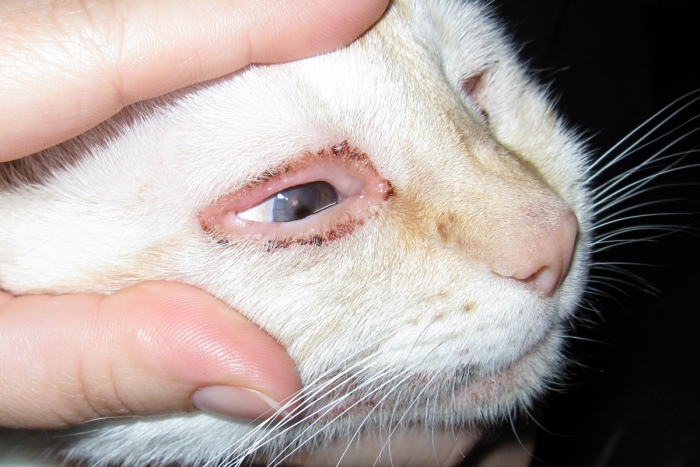
- Inflammation of the mucous membrane of the eye - ... The disease manifests itself as reddening of the conjunctiva, sometimes they turn red with a brown or bluish tinge. Also conjunctivitis causes photophobia, itching in the eye sockets, profuse or discharge from the eyes. In this case, the mucous membrane of the conjunctiva has a different degree of damage (wounds or ulcers).
- Increased discharge of tears - epiphora... This pathology is usually the result of an allergy to any irritant or an instinctive reaction aimed at prompt elimination foreign bodypenetrated into the visual organ. In pathological cases, epiphora occurs with obstruction of the lacrimal duct.
- Inflammation of the choroid - uveitis... The disease develops as a result of various infectious and (and) invasive infections, characterized by outflows of different consistency and color.
- Corneal inflammation - keratitis... For him, viscous yellowish or greenish outflows are indicative. At the same time, the eyes flow, the eyelids often stick together, overgrow with crusts.
Factors causing pathology
Since several diseases can lead to purulent discharge from the eyes, there are a lot of causes:
- infectious diseases. In particular, rhinotracheitis, panleukopenia, calcivirosis, chlamydia and others;
- invasive diseases. Toxoplasmosis is most often found, but other helminthiases are also possible;
- for dust, pollen, chemical vapors;
- chronic non-infectious pathologies: diabetes mellitus, damage to the liver, kidneys, digestive organs;
- mechanical damage;
- hair penetration into the eye (recorded in long-haired pets);
- breed predisposition;
- old age.
Diagnostics
The veterinarian needs to determine the causes of the disease based on the data of the initial examination and laboratory results.
If the discharge from the eyes is brown, yellow or green, then this indicates a bacterial infection.
It is especially important for the doctor to obtain the following information:
- At what point did the discharge appear.
- Exudate parameters (transparency, color, viscosity, volume and frequency of discharge).
- Injuries to or around the eye.
- When deworming activities were carried out.
- Than the pet has been ill lately.
To determine the pathogen, it is necessary to pass tests (exudate from the eyes). Medical history is of great importance in determining the diagnosis. Any owner must have a passport for his pet, in which everything that happens is recorded by the attending physician.
Types of exudate
The exudate may differ in color and consistency, which says a lot about the causes of the disease:
- Purulent exudate It comes in various shades, usually from white to yellow, but sometimes green or brown exudates are also found. Sometimes the pus hardens, and hard suppurations often remain color.
- Brown liquid discharge talk about epiphora - blockage of the nasolacrimal canal. This condition can be distinguished by expiration. They are more liquid, watery (thick pus) and at the same time abundant, observed in the corners of the eyes.
- Red-brown exudate is also purulent. It is obtained when many erythrocytes accumulate in the pus, which indicates a violation of the permeability of the capillaries of the conjunctiva or its mechanical damage.
Treatment of the disease
Sometimes it takes time to make an accurate diagnosis (for example, seeding of microflora from the eyes is needed). But treatment must be started immediately so that complications do not appear. When the diagnosis is clarified, the therapy is changed. First of all, an eye wash is prescribed to alleviate the condition of the pet, the owner acquires the drops based on the doctor's prescription.
To cleanse the eye, you need cotton pads or sterile gauze wipes, a disinfectant or other medication prescribed by your doctor, as well as a treat for the kitten (to reward him after unpleasant procedures). The owner of the animal can do such procedures at home, since the procedure is simple.

Cleansing the eyes of an animal is carried out in several stages:
- The animal is securely fixed; at home, this will require the help of a second person.
- Wipes or cotton pads are moistened with a medicinal or disinfectant solution.
- With one hand, you need to gently open your eyelids.
- With the other hand, also gently remove debris and exudate from the eyes, moving to the inner corner of the eye from the outside.
- After the procedure, put a healing ointment under the sick eyelid of the pet.
- So that the pet is not afraid of the next procedures, it is stroked, treated with delicacies.
Physiotherapy (warming up) is sometimes prescribed. In some cases, surgical intervention is required. In case of bacterial infection, you should rinse your eyes with antibacterial agents. Systemic antibiotics may also be prescribed, which must be given intramuscularly or given orally with food. The duration of the course of treatment is determined and prescribed by the doctor, usually with a bacterial infection, from 5-7 to 14-20 days is enough, it all depends on the type of pathogen.
Prevention
So that the pet does not suffer in the future, you need to follow some rules:
- Feed your pet with vitamins regularly
- Maintain hygiene (clean pot and bowl).
- Carry out deworming annually.
- Discourage communication with stray animals.
Discharge from a pet's eyes is the norm if they are transparent and there are few of them. You should be alert if you find black discharge from the eyes of a cat.
A clear liquid on the eyes can appear as a result of a reaction that protects the animal's eyes from environmental factors (wind, dust). The same reaction can occur if an irritant gets into the pet's nose. In Persian cats, lacrimation is noted after sleep. This is due to the structural features of their head. That is, if the animal feels good and the fluid secreted from the eyes is transparent, then the cat is healthy. If it has acquired a certain shade, then it's time to start worrying.
Causes of black discharge from cat eyes
If an adult cat has black discharge in front of his eyes, this may indicate the occurrence of various diseases in her. There are several reasons for such deviations.:
- Respiratory infection.
- Herpesvirus.
- Chlamydia.
Injury to the eye can also cause black discharge. Without proper treatment, the eyes may fester. The reason for this is infection. If the secreted substance turns brown, then the cat has an obstruction of the nasolacrimal canal.
Is black discharge from the eyes dangerous in cats?
By itself, the black discharge from the eyes of a cat does not pose any danger and does not affect its vision. However, they become a signal that your pet may have health problems. Despite the fact that the animal may be in its usual state, you need to seek help from a veterinary center. In order to make a diagnosis, the veterinarian will order an eye wash test.
Diagnostics includes a visual examination of the animal. In this case, mechanical tissue damage may be detected. If a part of the optic organ such as the cornea is damaged, the discharge will be brown. The animal may feel discomfort or pain. This is easy to see from his behavior. Anxiety, frequent blinking or squinting of the injured eye is a reason to immediately contact your veterinarian.
How to cure a cat
In addition to treating the underlying disease, the symptom of which was the increased secretion of the lacrimal glands and a change in their color, attention should be paid to the hygiene of the visual organs. You can remove black discharge from a cat with solutions of furacilin or boric acid. They will not only cleanse dirt, but also have an antibacterial effect. Solutions can be replaced with strong tea, chamomile broth, or just warm water. In some cases, the cat is prescribed antibacterial drops.

Instructions for the use of solutions
So that the animal does not get hurt and the procedure has an effect, you need to take a responsible approach to washing the eyes. To do this, you need to adhere to the following rules:
- Ask someone for help... Have one person wash the eyes while the other holds the animal tightly.
- Take a clean cotton swab... Wet it with solution, decoction or water.
- If your pet's eyelids are stuck together, carefully moisten them with a solution beforehand until they open.
- Blot stuck eyelashes with a damp swab from the nose to the corners of the eyes.
- Squeeze out the liquidtyped in a tampon into the cat's eye.
Always keep the tampons wet. Dry cotton wool can further injure the damaged eye. It is better to use warm solutions.
Please note that if your pet has black discharge in the eyes, you do not need to self-medicate. You can harm your cat's health. See your veterinarian promptly for the correct diagnosis and treatment.
Any experienced breeder knows very well that a cat's health can be judged with confidence by its appearance... If the pet is disheveled, its coat is dry and lifeless, and the look is sluggish and clouded, there is no need to talk about "heroic" health. In addition, brown discharge in cats from the eyes itself often indicates the presence of dangerous pathologies in the animal. The sooner you take your pet to the vet, the more likely the specialist will be able to help him.
General information
It is important to understand that almost everyone, as or otherwise affecting the condition of the eyes, can lead to the appearance of brown exudate. This symptom is not a characteristic symptom: any of hundreds of systemic pathologies can cause this phenomenon with equal probability.
In the best case, the appearance of a brownish exudate is a sign of debris getting into the eyes with subsequent development. That is why high-quality, timely diagnostics plays a very important role.
The pet owner himself can help with this. His task is to closely monitor the pet. It is necessary to notice in a timely manner signs indicating a deterioration of the pathological process, promptly informing the veterinarian about this. Insignificant amounts of brownish discharge in the corners of the eyes indicate a working treatment and the normal state of a sick animal, otherwise it is necessary to consult a specialist as soon as possible. Therapy depends on the identified underlying cause of the inflammation. We will talk about some of them below.
Predisposing factors
In veterinary practice, the following reasons are more often revealed, under the influence of which a brownish exudate can be released from the cat's eyes:
- Congenital or acquired disorders of eyelash growth. So, in some animals, eyelashes from birth do not grow outward, but inward. There are also rare cases when wool begins to break through the entire inner surface of the eyelids. Of course, in such animals the cornea is constantly wounded and damaged, develops chronic inflammation, which is accompanied by the release of large volumes of exudate.

- Acquired or congenital anomalies anatomical structure century. The most typical is them. The result is the same as in the previous case. The eyelashes are in direct contact with the surface of the cornea.
- Inflammation of the eyelids () or inflammation of the Meibomian glands. As we already said, any systemic infection can lead to this result.
- Prolapse (bulging) of the lacrimal gland. The pathology is also known as cherry eye.
- Benign tumors and oncological diseases.
- third century.
- Congenital and acquired anatomical defects of the lacrimal ducts. If they are clogged or pinched, the surface of the eyeball is not wetted by the tear. This leads to its drying out and the development of pathogenic (or conditionally pathogenic) microflora.
- . They are quite common in dogs. Even if this pathology is suspected, it is necessary to urgently visit the veterinarian, since in advanced cases it is necessary to remove the entire eyeball.
- All forms, as well as trauma to the conjunctival membrane itself and eyelids.
Of course, this is not all possible reasons... Below we list not so typical cases encountered, however, in the world veterinary practice:
- Eyes "flow" due to abrasions, scratches and other damage to the cornea.
- Injuries to the palate or bones of the facial skull.
- Inflammation of the cornea (keratitis).
- Congenital defects of the cornea and conjunctiva. In dogs, in particular, dermoids are often detected. This is the name of benign cystic formations, which contain epithelial mass, sebum, hair. It happens that they are hard to the touch, but more often they reveal elastic or "plastic" neoplasms.
- ... This is an inflammation of the choroid of the eyeball. It is rare in dogs.
- ... Pathology is accompanied not only by inflammation, exudate and soreness, but also sharp rise intraocular pressure, which under certain conditions can lead to the need for surgical removal of the eyeball.
- Lens pathology.
- Chronic periodontal disease. In such cases, brown discharge develops due to an infection that has risen through the nasolacrimal canal into the conjunctival cavity.
Diagnostic techniques
Since all eye diseases are potentially fraught with full or partial, the veterinarian, using the diagnostic algorithm below, seeks to find out as soon as possible about the root cause of the pathology:
- Complete medical history and physical examination.
- The Schirmer test is mandatory. It helps to identify the initial stages of keratoconjunctivitis. To detect surface defects, ulcers and corneal erosion, the application of the latest fluorescent formulations to the surface is used. Also shown is tonometry to determine intraocular pressure. If possible, all of these checks should be performed by a veterinary ophthalmologist who can accurately interpret the test results.

- Cytological examination of scrapings from the cornea and conjunctival cavity.
- Conducting complete blood tests and serological tests. These techniques allow the detection of systemic infections, provided they are present in the animal.
In doubtful and difficult cases, when the above methods did not allow obtaining an unambiguous result, you should use narrower diagnostic methods:
- Cultivation of a culture of the pathogen on nutrient media.
- Carrying out PCR reactions, as well as ELISA. They help to make an accurate diagnosis in case of suspected viral infections.
- Microscopic examination of scrapings from the cornea and conjunctiva.
- Collection of aspirates from neoplasms, if any, near the animal's eyes.
- Deep endoscopy of the nasal cavity is helpful. We have already said that infections from the sinuses of the skull can "make their way" into the eyes, so it will not be superfluous to examine them.
- Ultrasonography of the eye and its surrounding soft tissues.
- X-rays of the skull to identify fractures, sinus diseases and bone tumors.
- Dacryocystorinography. A method used to assess the condition of the tear ducts.
- In elusive ideal cases, MRI is done.
Treatment
The treatment prescribed depends on the underlying cause identified. So, in case of systemic infections, the problem is dealt with by injecting the animal with a wide spectrum of action and other antimicrobial drugs. Tumors, dermoids, twists of the eyelids, etc., are eliminated by resorting to surgery.
The owner himself can also help his pet. To do this, it is necessary to regularly remove the released exudate using a cotton pad dipped in warmth. saline... This should be done as often as possible, preventing the exudate from drying out with the subsequent formation of hard crusts. Also, it will not hurt to lay a tetracycline ointment into the conjunctival cavity, since this drug removes inflammation and promotes accelerated regeneration of the affected organ.